## Diagram: Line Drawings of Biological Structures
### Overview
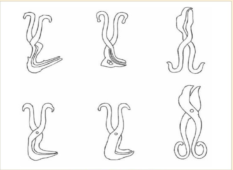
The image displays six distinct black-and-white line drawings arranged in a 2x3 grid (two rows, three columns) on a plain white background. The drawings appear to be technical or anatomical illustrations of a similar biological structure, possibly representing different developmental stages, variations, or perspectives of an organ or organism. There is no textual information, labels, axes, legends, or numerical data present in the image.
### Components/Axes
* **Layout:** A grid of six individual illustrations.
* **Content:** Each drawing consists of clean, continuous black lines forming a complex, organic shape. The shapes generally feature a central, somewhat bulbous or elongated body from which two symmetrical, curved appendages extend upwards. The lower portion of each structure varies, showing different configurations of folds, extensions, or coiled forms.
* **Notable Features:** Several drawings contain small, distinct circles or dots within the main body, which may represent specific anatomical landmarks (e.g., openings, nuclei, or points of interest).
### Detailed Analysis
**Spatial Grounding & Component Isolation:**
* **Top Row, Left:** Structure with two upward-curving appendages. The lower section features a prominent, forward-pointing, beak-like extension composed of multiple layered lines.
* **Top Row, Center:** Similar upward appendages. The lower section is more compact and rounded, with internal lines suggesting folds or layers. A small, irregular shape is visible within the lower body.
* **Top Row, Right:** The appendages are more elongated and wavy. The lower section splits into two distinct, downward-curving hooks or tendrils.
* **Bottom Row, Left:** Upward appendages are present. The lower body is a single, smooth, curved form pointing to the right. A small circle is located centrally within the main body.
* **Bottom Row, Center:** Very similar to the bottom-left drawing, with upward appendages and a smooth, right-pointing lower curve. It also contains a central small circle.
* **Bottom Row, Right:** The most distinct variation. The upward appendages are replaced by two large, leaf-like or petal-like forms. The lower section consists of two symmetrical, tightly coiled spirals. A small circle is present in the central body.
### Key Observations
1. **Symmetry and Variation:** All structures exhibit a degree of bilateral symmetry in their upper appendages. The primary variation occurs in the morphology of the lower section.
2. **Recurring Motif:** The small circle appears in three of the six drawings (bottom-left, bottom-center, bottom-right), suggesting it may be a key feature being highlighted in those specific variants.
3. **Stylistic Consistency:** The uniform line weight and lack of shading indicate these are schematic diagrams meant to convey form and structure rather than realistic texture or depth.
### Interpretation
This image is a **comparative morphological chart**. Its purpose is to visually catalog and contrast different forms of a single biological entity. The absence of text implies it is meant to accompany a descriptive text or be part of a series where the labels are provided separately.
* **What the data suggests:** The diagrams likely illustrate evolutionary adaptations, developmental stages (ontogeny), or taxonomic variations within a species or related group. The progression from left to right, top to bottom, may show a sequence, but without labels, this is speculative.
* **How elements relate:** The grid layout facilitates direct visual comparison. The viewer's eye is drawn to compare the consistent upper structure against the highly variable lower structure, emphasizing that the lower region is the primary site of morphological difference.
* **Notable patterns/anomalies:** The bottom-right drawing is the most divergent, breaking the pattern of simple upward appendages with its large, leaf-like structures and coiled base. This could represent a specialized form, a different viewing angle, or a distinct developmental endpoint. The pairing of the nearly identical bottom-left and bottom-center drawings suggests they may represent a standard form or a control against which others are compared.
**Conclusion:** This is a technical illustration designed for visual analysis in a biological or medical context. To extract its full informational value, one would need the accompanying figure legend or caption, which is not present in the image itself. The image provides **structural and comparative data** but no quantitative or labeled factual data.