## Diagram: Plastic Repair of Ear Lobe
### Overview
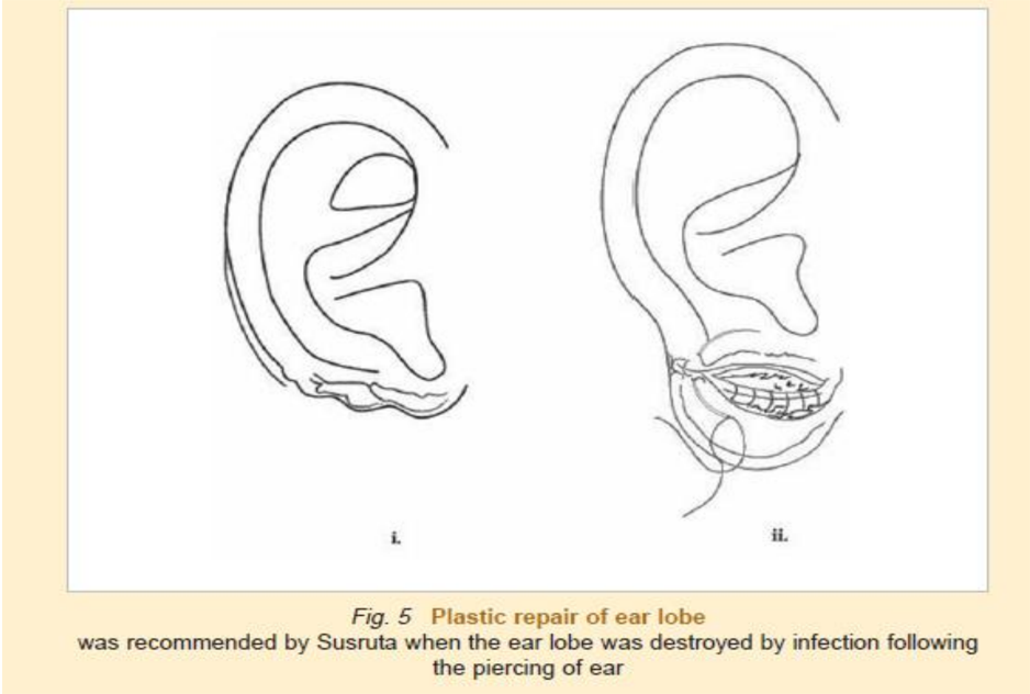
The image is a black-and-white technical diagram illustrating two stages of ear lobe repair. It includes labeled anatomical sketches and explanatory text.
### Components/Axes
- **Left Illustration (i.)**: A normal, intact ear lobe with defined anatomical contours (helix, antihelix, lobule).
- **Right Illustration (ii.)**: A repaired ear lobe showing a suture line (dashed line) extending from the base of the lobe to the earlobe margin.
- **Text Elements**:
- Title: "Plastic repair of ear lobe" (bold, centered).
- Caption: "was recommended by Susruta when the ear lobe was destroyed by infection following the piercing of ear" (italicized, centered).
- **Labels**:
- "i." (below left illustration).
- "ii." (below right illustration).
### Detailed Analysis
- **Left Illustration (i.)**:
- Depicts a standard ear lobe with no visible damage or repair.
- Anatomical features include the outer rim (helix), inner curvature (antihelix), and lobule.
- **Right Illustration (ii.)**:
- Shows a repaired ear lobe with a suture line (dashed line) bridging the gap caused by tissue loss.
- The suture extends from the base of the lobe (near the face) to the earlobe margin, suggesting a tension-free closure.
- **Textual Context**:
- The caption references Susruta, an ancient Indian surgeon, indicating historical precedent for the repair technique.
- The repair is described as addressing tissue destruction from infection post-piercing.
### Key Observations
- The diagram emphasizes the transition from a damaged ear lobe (implied by the caption) to a repaired state via suturing.
- No numerical data or quantitative values are present; the focus is on anatomical and procedural representation.
- The suture line in (ii.) is the only explicit indication of the repair method.
### Interpretation
- The diagram serves as a visual guide for surgical techniques in ear lobe reconstruction, highlighting the importance of tension-free closure to prevent recurrence of infection or deformity.
- The reference to Susruta underscores the longevity of this approach, suggesting its validation in historical medical practices.
- The absence of color or additional annotations limits the ability to infer material properties (e.g., suture type) or patient-specific variables.
- The simplicity of the diagram prioritizes clarity over detail, making it suitable for educational or procedural reference.