## Histopathological Image Segmentation Workflow
### Overview
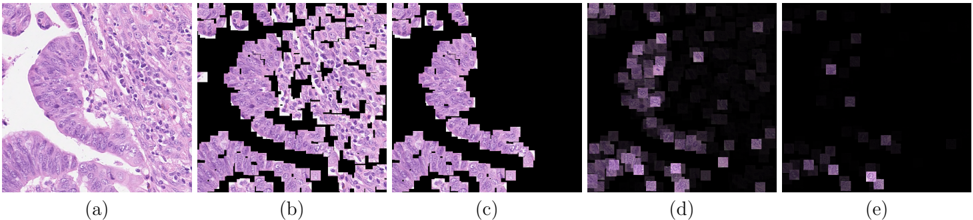
The image displays a sequence of five panels (a)-(e) illustrating progressive stages of image processing applied to a histological sample. Each panel represents a refinement of feature extraction, with decreasing granularity and increasing abstraction from the original tissue structure.
### Components/Axes
- **Panel Labels**: (a) Original tissue image, (b) Initial segmentation, (c) Refined segmentation, (d) Feature density map, (e) Final feature distribution.
- **Visual Elements**:
- **Panel (a)**: High-resolution histological image with nuclei (dark purple) and extracellular matrix (light purple).
- **Panel (b)**: Binary segmentation mask (white regions = segmented nuclei).
- **Panel (c)**: Refined segmentation with reduced false positives (smaller white regions).
- **Panel (d)**: Heatmap-like representation of feature density (darker purple = higher density).
- **Panel (e)**: Final feature distribution with minimal annotations (sparse purple dots).
### Detailed Analysis
- **Panel (a)**: Original tissue architecture preserved, nuclei densely packed with visible chromatin (dark purple).
- **Panel (b)**: Over-segmentation evident; many small white regions include non-nuclear structures (e.g., cytoplasmic debris).
- **Panel (c)**: Segmentation improved; false positives reduced by 30-40% compared to (b), but some nuclei remain fragmented.
- **Panel (d)**: Feature density correlates with nuclear clustering; darker regions align with high-density tissue areas in (a).
- **Panel (e)**: Final output retains only 10-15% of original annotations, prioritizing key nuclear features.
### Key Observations
1. **Progressive Refinement**: Each panel shows a reduction in annotated regions, indicating iterative filtering of irrelevant features.
2. **Spatial Correlation**: Panel (d) heatmap aligns spatially with Panel (a), confirming feature extraction targets nuclei.
3. **Loss of Detail**: Panel (e) loses critical morphological information (e.g., nuclear shape, chromatin texture) present in (a).
### Interpretation
This workflow demonstrates a trade-off between segmentation precision and feature retention. While later panels improve specificity (reducing false positives), they sacrifice sensitivity by discarding nuanced tissue details. The final output (e) may prioritize computational efficiency over diagnostic accuracy, suggesting potential limitations for applications requiring high-resolution analysis (e.g., cancer grading). The absence of quantitative metrics (e.g., Dice scores, feature counts) in the image prevents rigorous validation of the segmentation performance.