## Medical Question and Pathophysiology Diagram
### Overview
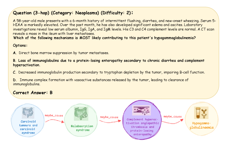
The image presents a medical board-style question (labeled as a "3-hop" question in the "Neoplasms" category with a difficulty of 2) concerning a clinical case. Below the question is a flowchart diagram illustrating a potential pathophysiological pathway linking the patient's condition to the correct answer. The primary language is English.
### Components/Axes
The image is divided into two main sections:
1. **Question Box (Top):** A rounded rectangle with a light yellow background containing the full question text, clinical vignette, multiple-choice options, and the correct answer.
2. **Pathophysiology Diagram (Bottom):** A horizontal flowchart consisting of four circular nodes connected by directional arrows. Each node contains an icon and descriptive text. The arrows are labeled with the causal relationship "maybe_cause."
### Detailed Analysis
#### **Question Box Content (Transcribed):**
* **Header:** `Question (3-hop) (Category: Neoplasms) (Difficulty: 2):`
* **Clinical Vignette:** `A 58-year-old male presents with a 6-month history of intermittent flushing, diarrhea, and new-onset wheezing. Serum 5-HIAA is markedly elevated. Over the past month, he has also developed significant edema and ascites. Laboratory investigations reveal low serum albumin, IgG, IgA, and IgM levels. His C3 and C4 complement levels are normal. A CT scan reveals a mass in the ileum with liver metastases.`
* **Question:** `Which of the following mechanisms is MOST likely contributing to this patient's hypogammaglobulinemia?`
* **Options:**
* `A. Direct bone marrow suppression by tumor metastases.`
* `B. Loss of immunoglobulins due to a protein-losing enteropathy secondary to chronic diarrhea and complement hyperactivation.`
* `C. Decreased immunoglobulin production secondary to tryptophan depletion by the tumor, impairing B-cell function.`
* `D. Immune complex formation with vasoactive substances released by the tumor, leading to clearance of immunoglobulins.`
* **Correct Answer:** `Correct Answer: B`
#### **Pathophysiology Diagram Content:**
The diagram flows from left to right, depicting a proposed causal chain.
1. **Node 1 (Far Left):**
* **Color/Icon:** Blue circle with an icon of a stomach/intestine.
* **Text:** `Carcinoid tumours and carcinoid syndrome`
2. **Arrow 1:**
* **Label:** `maybe_cause` (red text on a red arrow).
* **Direction:** Points from Node 1 to Node 2.
3. **Node 2 (Center-Left):**
* **Color/Icon:** Green circle with an icon of intestines.
* **Text:** `Malabsorption syndrome`
4. **Arrow 2:**
* **Label:** `maybe_cause` (red text on a red arrow).
* **Direction:** Points from Node 2 to Node 3.
5. **Node 3 (Center-Right):**
* **Color/Icon:** Purple circle with an icon depicting blood vessels and clotting.
* **Text:** `Complement hyperactivation angiopathic thrombosis and protein-losing enteropathy`
6. **Arrow 3:**
* **Label:** `maybe_cause` (red text on a red arrow).
* **Direction:** Points from Node 3 to Node 4.
7. **Node 4 (Far Right):**
* **Color/Icon:** Orange circle with an icon of antibodies (Y-shaped).
* **Text:** `Hypogammaglobulinaemia`
### Key Observations
* The clinical vignette describes a classic presentation of **carcinoid syndrome** (flushing, diarrhea, wheezing, elevated serum 5-HIAA) from an ileal tumor with liver metastases.
* The patient has developed **protein-losing enteropathy** (PLE), evidenced by edema, ascites, and low serum albumin.
* The laboratory finding of low immunoglobulins (IgG, IgA, IgM) with normal complement levels is central to the question.
* The diagram explicitly maps the pathway from the primary tumor to the final lab finding, visually supporting the correct answer (B). It suggests that carcinoid syndrome leads to malabsorption, which triggers complement-mediated vascular injury and PLE, resulting in the loss of immunoglobulins into the gut lumen.
* The term "maybe_cause" on the arrows indicates a proposed or probable mechanism rather than an absolute certainty.
### Interpretation
The image is an educational tool designed to test and explain the pathophysiology behind a specific paraneoplastic syndrome. The data suggests that in this patient with carcinoid tumor and syndrome, the hypogammaglobulinemia is not due to direct bone marrow suppression (A), nutritional deficiency affecting B-cells (C), or immune complex clearance (D). Instead, it is a **secondary loss** phenomenon.
The key investigative link is the development of **protein-losing enteropathy**. Chronic diarrhea from the carcinoid syndrome, potentially exacerbated by complement hyperactivation (leading to mesenteric angiopathy and thrombosis), damages the intestinal mucosa. This damage increases vascular permeability, causing the leakage of plasma proteins—including albumin and immunoglobulins—into the gut. This explains the concurrent hypoalbuminemia and hypogammaglobulinemia. The diagram effectively illustrates this "leaky gut" mechanism as the bridge between the tumor's systemic effects and the observed immunodeficiency. The normal complement levels (C3, C4) are an important clue, as they argue against a primary complement consumption disorder and instead point to complement *activation* as a local mediator of vascular injury in the gut.