## Medical Diagram: Hypogammaglobulinemia Mechanisms
### Overview
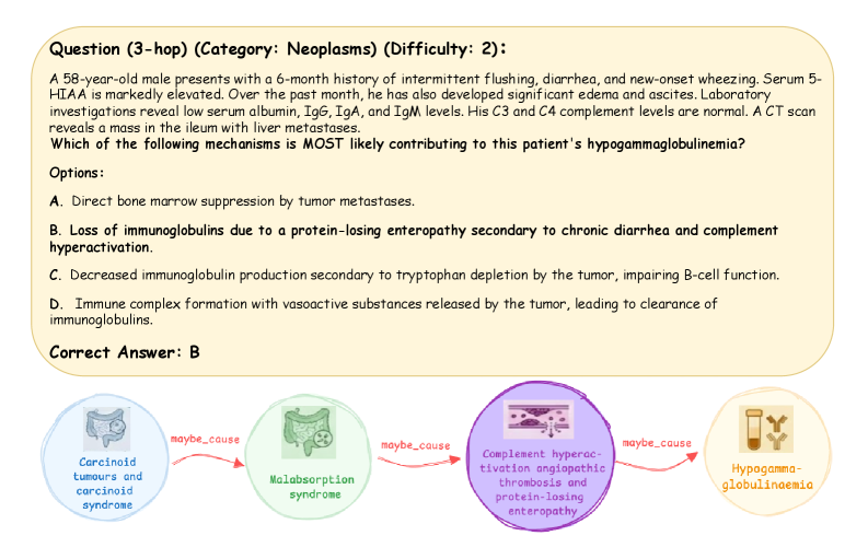
The image presents a medical question and a diagram illustrating potential mechanisms leading to hypogammaglobulinemia. The question describes a patient case, followed by multiple-choice options and the correct answer. The diagram visually connects "Carcinoid tumours and carcinoid syndrome" to "Hypogamma-globulinaemia" through intermediate steps like "Malabsorption syndrome" and "Complement hyperactivation angiopathic thrombosis and protein-losing enteropathy," suggesting a possible causal pathway.
### Components/Axes
* **Question Text:** Describes a patient case with symptoms and lab results.
* Category: Neoplasms
* Difficulty: 2
* **Options (A-D):** Multiple-choice answers presenting different potential mechanisms.
* **Correct Answer:** B
* **Diagram:** A flow diagram showing a potential causal pathway.
* Nodes:
* Carcinoid tumours and carcinoid syndrome (top-left, light blue circle)
* Malabsorption syndrome (center-left, light green circle)
* Complement hyperactivation angiopathic thrombosis and protein-losing enteropathy (center-right, purple circle)
* Hypogamma-globulinaemia (bottom-right, light brown circle)
* Edges: Arrows connecting the nodes, labeled "maybe_cause".
### Detailed Analysis or Content Details
* **Question:** A 58-year-old male presents with a 6-month history of intermittent flushing, diarrhea, and new-onset wheezing. Serum 5-HIAA is markedly elevated. Over the past month, he has also developed significant edema and ascites. Laboratory investigations reveal low serum albumin, IgG, IgA, and IgM levels. His C3 and C4 complement levels are normal. A CT scan reveals a mass in the ileum with liver metastases. Which of the following mechanisms is MOST likely contributing to this patient's hypogammaglobulinemia?
* **Options:**
* A. Direct bone marrow suppression by tumor metastases.
* B. Loss of immunoglobulins due to a protein-losing enteropathy secondary to chronic diarrhea and complement hyperactivation.
* C. Decreased immunoglobulin production secondary to tryptophan depletion by the tumor, impairing B-cell function.
* D. Immune complex formation with vasoactive substances released by the tumor, leading to clearance of immunoglobulins.
* **Correct Answer:** B
* **Diagram Nodes:**
* **Carcinoid tumours and carcinoid syndrome:** Located in the top-left, represented by a light blue circle containing a simplified illustration of the digestive system.
* **Malabsorption syndrome:** Located in the center-left, represented by a light green circle containing a simplified illustration of the digestive system.
* **Complement hyperactivation angiopathic thrombosis and protein-losing enteropathy:** Located in the center-right, represented by a purple circle containing a simplified illustration of blood vessels.
* **Hypogamma-globulinaemia:** Located in the bottom-right, represented by a light brown circle containing a simplified illustration of a test tube with antibodies.
* **Diagram Edges:**
* An arrow labeled "maybe_cause" connects "Carcinoid tumours and carcinoid syndrome" to "Malabsorption syndrome."
* An arrow labeled "maybe_cause" connects "Malabsorption syndrome" to "Complement hyperactivation angiopathic thrombosis and protein-losing enteropathy."
* An arrow labeled "maybe_cause" connects "Complement hyperactivation angiopathic thrombosis and protein-losing enteropathy" to "Hypogamma-globulinaemia."
### Key Observations
* The question presents a complex clinical scenario requiring knowledge of neoplasm-related complications.
* The diagram simplifies a potential pathophysiological pathway, highlighting the interconnectedness of different medical conditions.
* The "maybe_cause" labels on the arrows indicate that the diagram represents a possible, but not definitive, causal relationship.
### Interpretation
The image combines a clinical question with a simplified diagram to illustrate a potential mechanism leading to hypogammaglobulinemia in a patient with carcinoid tumors. The diagram suggests that carcinoid tumors may lead to malabsorption syndrome, which in turn may cause complement hyperactivation, angiopathic thrombosis, and protein-losing enteropathy. This ultimately may result in hypogammaglobulinemia. The question tests the understanding of these complex relationships and the ability to identify the most likely contributing mechanism based on the patient's presentation. The diagram serves as a visual aid to understand the potential causal chain, emphasizing the importance of considering multiple factors in diagnosing and treating medical conditions.