## 2D Contour Plot with Marginal Distributions: Text Type Clustering
### Overview
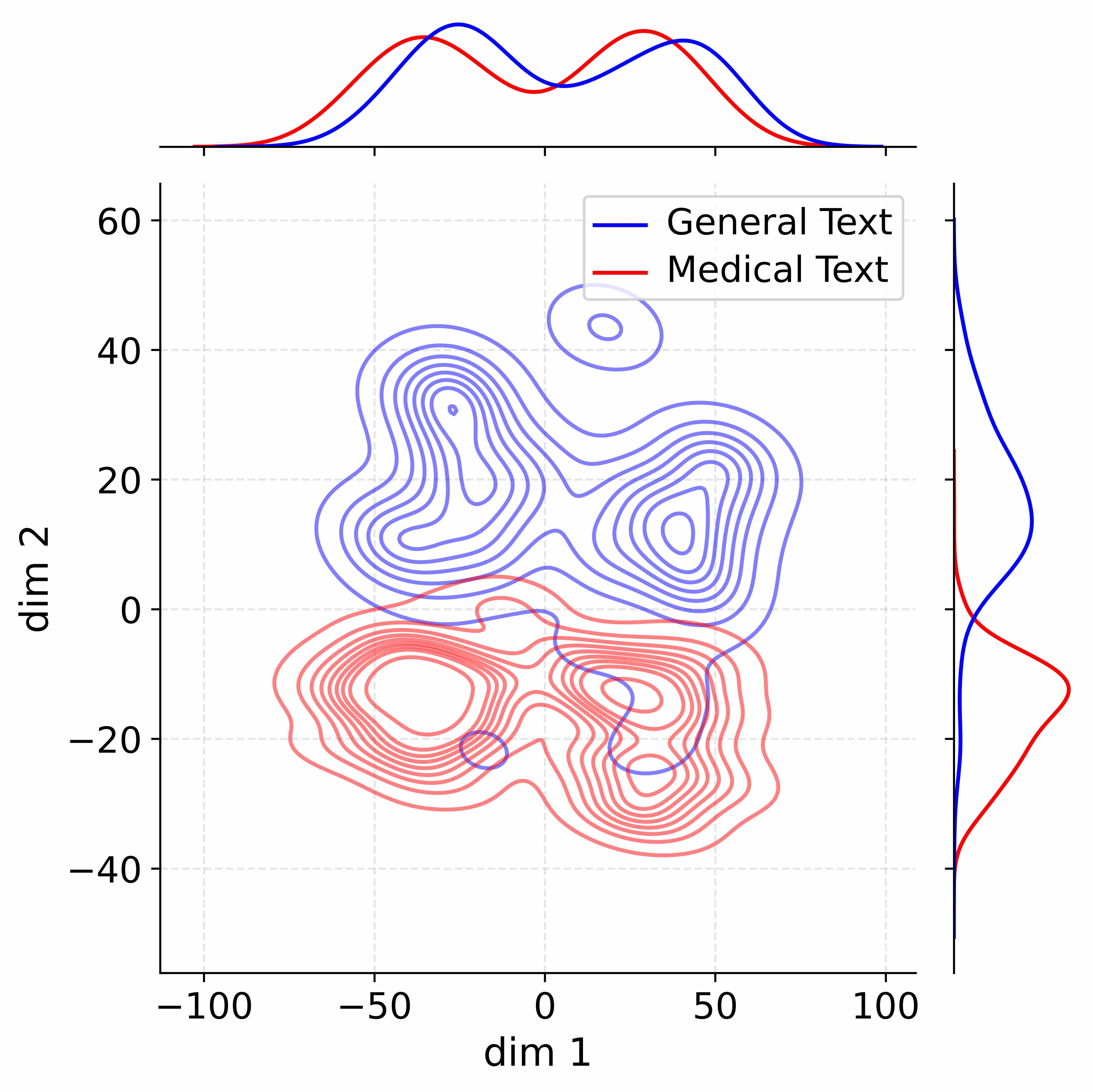
The image is a statistical visualization comparing the distribution of two text corpora ("General Text" and "Medical Text") in a two-dimensional latent space. It consists of a central 2D contour plot showing the joint density of the data points, accompanied by marginal distribution plots (1D density curves) along the top (for dim 1) and right side (for dim 2). The plot suggests the data has been projected or embedded into two dimensions, likely via a technique like PCA, t-SNE, or UMAP.
### Components/Axes
* **Main Plot (Center):**
* **X-axis:** Labeled "dim 1". Scale ranges from -100 to 100, with major tick marks at -100, -50, 0, 50, 100.
* **Y-axis:** Labeled "dim 2". Scale ranges from -40 to 60, with major tick marks at -40, -20, 0, 20, 40, 60.
* **Content:** Overlaid contour plots for two categories.
* **Legend:** Positioned in the top-right quadrant of the main plot area.
* A blue line corresponds to the label "General Text".
* A red line corresponds to the label "Medical Text".
* **Marginal Distribution Plots:**
* **Top Plot:** Shows the 1D density distribution along "dim 1". The x-axis aligns with the main plot's x-axis (-100 to 100). The y-axis represents probability density (unlabeled).
* **Right Plot:** Shows the 1D density distribution along "dim 2". The y-axis aligns with the main plot's y-axis (-40 to 60). The x-axis represents probability density (unlabeled).
### Detailed Analysis
**1. Main Contour Plot (Joint Distribution):**
* **General Text (Blue Contours):**
* **Spatial Grounding:** Primarily occupies the upper half of the plot (positive "dim 2" values).
* **Trend/Shape:** Forms a complex, multi-modal distribution. The highest density region (innermost contours) is centered approximately at (dim1 ≈ -25, dim2 ≈ 30). A secondary, smaller high-density cluster is visible around (dim1 ≈ 25, dim2 ≈ 15). A distinct, isolated small cluster appears at approximately (dim1 ≈ 15, dim2 ≈ 45). The overall spread spans dim1 from roughly -75 to 75 and dim2 from 0 to 50.
* **Medical Text (Red Contours):**
* **Spatial Grounding:** Primarily occupies the lower half of the plot (negative "dim 2" values).
* **Trend/Shape:** Also forms a multi-modal distribution. The highest density region is centered around (dim1 ≈ -25, dim2 ≈ -15). Another significant high-density cluster is located at approximately (dim1 ≈ 25, dim2 ≈ -25). The overall spread spans dim1 from roughly -75 to 75 and dim2 from -40 to 0.
* **Overlap:** There is a region of moderate overlap between the two distributions along the "dim 1" axis, particularly between dim1 values of -25 and 25, and around dim2 = 0. However, their primary densities are clearly separated along the "dim 2" axis.
**2. Marginal Distribution Plots:**
* **Top Marginal (dim 1):**
* **General Text (Blue):** Shows a bimodal distribution. Peaks are located at approximately dim1 = -30 and dim1 = 30. The valley between peaks is near dim1 = 0.
* **Medical Text (Red):** Also shows a bimodal distribution. Peaks are located at approximately dim1 = -35 and dim1 = 25. The valley is near dim1 = -5.
* **Comparison:** The distributions along dim 1 are similar in shape and range for both text types, with significant overlap. The Medical Text peaks appear slightly shifted left compared to the General Text peaks.
* **Right Marginal (dim 2):**
* **General Text (Blue):** Shows a unimodal distribution with a peak at approximately dim2 = 25. The distribution is skewed, with a longer tail extending towards lower dim2 values.
* **Medical Text (Red):** Shows a unimodal distribution with a peak at approximately dim2 = -20. The distribution is also skewed, with a longer tail extending towards higher dim2 values.
* **Comparison:** This plot reveals the most significant separation. The two distributions have distinct peaks on opposite sides of dim2 = 0, with minimal overlap. This confirms that "dim 2" is the primary axis differentiating the two text types.
### Key Observations
1. **Clear Separation on dim 2:** The most prominent feature is the distinct separation of the two text corpora along the "dim 2" axis. General Text clusters in the positive region, Medical Text in the negative region.
2. **Similarity on dim 1:** Both text types exhibit similar, bimodal distributions along "dim 1", suggesting this dimension captures a common structural variation present in both general and medical language.
3. **Multi-modal Structure:** Both distributions in the 2D space are multi-modal, indicating that each text type likely contains several distinct sub-categories or topics within the analyzed corpus.
4. **Isolated Cluster:** The small, isolated blue cluster at (15, 45) represents a subset of General Text that is distinct from the main body of general text in this latent space.
### Interpretation
This visualization demonstrates that when text data (likely from embeddings or feature vectors) is reduced to two dimensions, **"General Text" and "Medical Text" form largely separable clusters.** The axis labeled "dim 2" appears to capture a semantic or stylistic feature that strongly differentiates medical discourse from general language. This could relate to vocabulary specificity, syntactic complexity, or topic focus inherent to medical literature.
The overlap along "dim 1" suggests that both text types share some underlying common structure or variability. The multi-modal nature of each cluster implies that neither "General Text" nor "Medical Text" is monolithic; each contains internal groupings, which could correspond to different genres (e.g., news vs. fiction for general; clinical reports vs. research articles for medical).
The plot provides strong visual evidence that a model or analysis using these two dimensions could effectively distinguish between general and medical text, with "dim 2" being the most discriminative feature. The isolated blue cluster is an outlier warranting further investigation to understand what specific subset of general text it represents.