\n
## Diagram: Neural Activity Visualization
### Overview
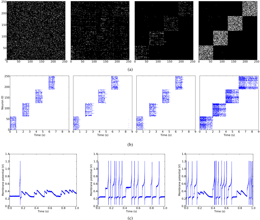
The image presents a series of visualizations depicting neural activity over time. It consists of three rows, labeled (a), (b), and (c). Row (a) shows spatial distributions of neuron activity as a binary pattern (presence/absence). Row (b) displays the activity of individual neurons (Neuron ID vs. Time) as a scatter plot. Row (c) shows the membrane potential of a subset of neurons over time as a line graph. The visualizations appear to be snapshots taken at different time points, illustrating the evolution of neural activity.
### Components/Axes
* **Row (a):** Four subplots showing spatial distributions. X-axis: Position (0-250, units unspecified). Y-axis: Position (0-250, units unspecified). The plots are binary, showing active (white) and inactive (black) neurons.
* **Row (b):** Four subplots showing Neuron ID vs. Time. X-axis: Time (0-9 seconds). Y-axis: Neuron ID (0-250). Points represent neuron activity at a given time.
* **Row (c):** Three subplots showing Membrane Potential vs. Time. X-axis: Time (0-1 second). Y-axis: Membrane Potential (0-1.4 Volts). Lines represent the membrane potential of neurons over time.
* **Labels:** (a), (b), (c) are labels for the rows.
### Detailed Analysis or Content Details
**Row (a):**
* **Plot 1:** Sparse activity, scattered white points across the space.
* **Plot 2:** Activity appears to be consolidating into a more defined region, roughly centered.
* **Plot 3:** The active region is more concentrated and elongated, extending diagonally.
* **Plot 4:** The active region has further consolidated into a more compact, diagonal band.
**Row (b):**
* **Plot 1:** Neurons exhibit relatively random activity across the time range. The points are scattered.
* **Plot 2:** A pattern begins to emerge, with some clustering of activity around Neuron IDs 50-150.
* **Plot 3:** The pattern becomes more pronounced, with clear vertical bands of activity at approximately Time = 2s, 5s, and 8s.
* **Plot 4:** The vertical bands are more defined, and there's some activity spreading between the bands.
**Row (c):**
* **Plot 1:** The membrane potential fluctuates around 0.2V, with occasional spikes up to approximately 0.6V. The spikes are relatively infrequent.
* **Plot 2:** The membrane potential shows more frequent spikes, reaching up to approximately 1.2V. The baseline potential appears slightly higher than in Plot 1.
* **Plot 3:** The membrane potential exhibits a series of rapid, high-amplitude spikes, reaching approximately 1.4V. The spikes are clustered together, followed by periods of lower activity.
### Key Observations
* The activity in Row (a) transitions from a dispersed state to a more localized and organized pattern.
* Row (b) shows a corresponding shift from random activity to a more structured, periodic pattern.
* Row (c) demonstrates an increase in the frequency and amplitude of membrane potential spikes over time.
* The patterns observed in all three rows appear to be correlated, suggesting a coordinated evolution of neural activity.
### Interpretation
The data suggests a progression of neural activity, potentially representing the initiation and propagation of a signal or wave. The initial dispersed activity (a, b, c - Plot 1) indicates a baseline level of neuronal firing. As time progresses, the activity becomes more focused spatially (a) and temporally (b), leading to increased membrane potential fluctuations and spiking (c). This could represent the recruitment of neurons into a network, the formation of a traveling wave, or the onset of a specific cognitive process. The increasing amplitude and frequency of spikes in Row (c) suggest that neurons are becoming more excitable and actively participating in the signal. The vertical bands in Row (b) indicate synchronized activity among a subset of neurons at specific time points. The correlation between the three rows suggests that the observed patterns are not random but rather reflect a coherent underlying process. The data does not provide information about the specific type of neural activity or the underlying biological mechanisms.