TECHNICAL ASSET FINGERPRINT
307c52fc5219d920013b5aee
Click to view fullscreen
Press ESC or click to close
FOUND IN PAPERS
EXPERT: healer-alpha-free VERSION 1
RUNTIME: free/openrouter/healer-alpha
INTEL_VERIFIED
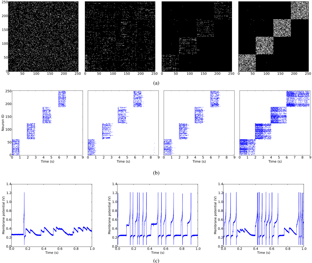
## Composite Neural Activity Figure: Network Connectivity, Spike Rasters, and Membrane Potentials
### Overview
This image is a three-part scientific figure, labeled (a), (b), and (c), illustrating the activity of a simulated neural network. It progresses from showing the network's structural connectivity (a), to its collective spiking activity over time (b), and finally to the detailed membrane potential dynamics of individual neurons (c). The figure demonstrates the emergence of structured, synchronized activity from an initially random state.
### Components/Axes
The figure is divided into three horizontal panels:
**Panel (a): Network Connectivity/Activity Matrices**
* **Type:** Four square raster plots (250x250 pixels/units).
* **Axes:** Both the X and Y axes are labeled from 0 to 250. No explicit axis titles are provided, but in this context, they typically represent neuron indices (e.g., pre-synaptic neuron ID on X, post-synaptic neuron ID on Y).
* **Content:** White pixels on a black background. The four plots show a clear progression from left to right.
* **Spatial Grounding:** The four subplots are arranged in a single row, labeled implicitly by their order.
**Panel (b): Network Spike Rasters**
* **Type:** Four line/scatter plots showing spike times.
* **Y-axis:** Labeled "Neuron ID", ranging from 0 to 250.
* **X-axis:** Labeled "Time (s)", ranging from 0 to 9.
* **Data Series:** Blue dots represent individual action potentials (spikes) fired by a specific neuron (Y-axis) at a specific time (X-axis).
* **Spatial Grounding:** The four subplots are arranged in a single row directly below panel (a), maintaining the same left-to-right progression.
**Panel (c): Single-Neuron Membrane Potentials**
* **Type:** Three line graphs.
* **Y-axis:** Labeled "Membrane potential (V)", ranging from 0.0 to 1.4.
* **X-axis:** Labeled "Time (s)", ranging from 0.0 to 1.0.
* **Data Series:** A continuous blue line traces the voltage of a single neuron over a 1-second window.
* **Spatial Grounding:** The three subplots are arranged in a single row at the bottom of the figure.
### Detailed Analysis
**Panel (a) - Connectivity/Activity Progression:**
1. **Leftmost Plot:** Appears as near-uniform random noise. White pixels are scattered densely and randomly across the entire 250x250 grid. This suggests an initial, unstructured state.
2. **Second Plot:** Faint, block-like structures begin to emerge along the diagonal. The background remains noisy, but clusters of higher pixel density are visible.
3. **Third Plot:** The diagonal block structure is much more pronounced. Four to five distinct, dense square blocks are visible along the main diagonal, with significantly less noise in the off-diagonal regions.
4. **Rightmost Plot:** Shows a very clear, sharp block-diagonal structure. Five dense, well-defined square blocks of white pixels sit on the main diagonal against a nearly black (empty) background. This indicates a highly segregated or modular network organization.
**Panel (b) - Spike Raster Progression:**
* **Trend Verification:** All four plots show a diagonal banding pattern, where groups of neurons (clusters on the Y-axis) fire in sequence over time (X-axis). The clarity and definition of these bands increase from left to right.
1. **Leftmost Plot:** Bands are diffuse and overlapping. Neuron clusters fire in broad, poorly timed windows (e.g., cluster ~0-50 fires from ~0-1s, cluster ~50-100 from ~1-2s, etc.).
2. **Second Plot:** Bands become slightly tighter and more separated in time.
3. **Third Plot:** Bands are well-defined and separated. Each cluster of neurons fires in a distinct, narrow time window with minimal overlap.
4. **Rightmost Plot:** Bands are extremely sharp and precise. The firing of each neuron cluster is tightly synchronized and confined to a very specific ~1-second interval, creating a perfect diagonal cascade.
**Panel (c) - Membrane Potential Dynamics:**
1. **Left Plot:** Shows a neuron with sparse, irregular spiking. The membrane potential hovers around a baseline of ~0.25V, with occasional sharp spikes reaching ~1.2V. Between spikes, there are small, irregular fluctuations.
2. **Middle Plot:** Shows a neuron with regular, periodic spiking. The baseline is stable at ~0.25V. Rhythmic spikes to ~1.2V occur at a steady frequency of approximately 10-12 Hz (10-12 spikes per second). There is a brief period of depolarization block (sustained higher voltage around 0.5V) between 0.4s and 0.5s.
3. **Right Plot:** Shows a neuron with burst-like spiking activity. The pattern is less regular than the middle plot. It features clusters of 2-4 rapid spikes followed by short pauses. The baseline potential shows more variability.
### Key Observations
1. **Structural-Functional Correlation:** There is a direct visual correlation between the emergence of block-diagonal structure in panel (a) and the emergence of precise, sequential cluster firing in panel (b).
2. **Temporal Precision:** The progression in panel (b) shows a dramatic increase in the temporal precision of neural population activity, moving from sloppy, overlapping bursts to a clean, sequential activation pattern.
3. **Diverse Single-Neuron Dynamics:** Panel (c) reveals that within this structured network activity, individual neurons can exhibit different intrinsic firing patterns (sparse, regular, bursting).
4. **Voltage Scale:** All membrane potential plots in (c) share the same Y-axis scale (0.0-1.4V), allowing direct comparison of spike amplitudes and baseline levels.
### Interpretation
This figure likely illustrates the results of a neural network simulation undergoing a process of **self-organization or learning**.
* **What the data suggests:** The progression from left to right across panels (a) and (b) demonstrates a network evolving from a random, unstructured state into a highly organized, modular state. The final state (rightmost plots) shows a network where distinct groups of neurons are connected within themselves (dense blocks in `a`) and fire in a precise, sequential order (clean diagonal in `b`). This is characteristic of a **synfire chain** or a **sequence-generating network**, where activity propagates reliably through a chain of neuronal groups.
* **How elements relate:** Panel (a) shows the underlying *structural* connectivity that enables the *functional* activity pattern seen in (b). The block-diagonal connectivity means neurons within a group are strongly connected, promoting synchronized firing within the group, while connections between groups are arranged to drive the sequential activation. Panel (c) provides a *biophysical* view, showing how individual neurons within such a network might behave, with their spiking patterns contributing to the population rhythm.
* **Notable anomalies:** The brief depolarization block in the middle plot of panel (c) is an interesting detail. It may represent a temporary saturation or inactivation of the neuron's spike-generating mechanism due to strong, rhythmic input from the synchronized network.
* **Underlying mechanism:** The transformation likely results from a **Hebbian-like learning rule** (e.g., spike-timing-dependent plasticity) applied to the network. Neurons that fire together (within a cluster) strengthen their connections, solidifying the blocks. The sequential activation is shaped by the initial random connections between clusters, which are selectively strengthened to create the forward-propagating sequence. The figure powerfully visualizes how local synaptic changes can give rise to global, ordered network dynamics.
DECODING INTELLIGENCE...