## Heatmap and Line Graph Analysis: Neuronal Activity Over Time
### Overview
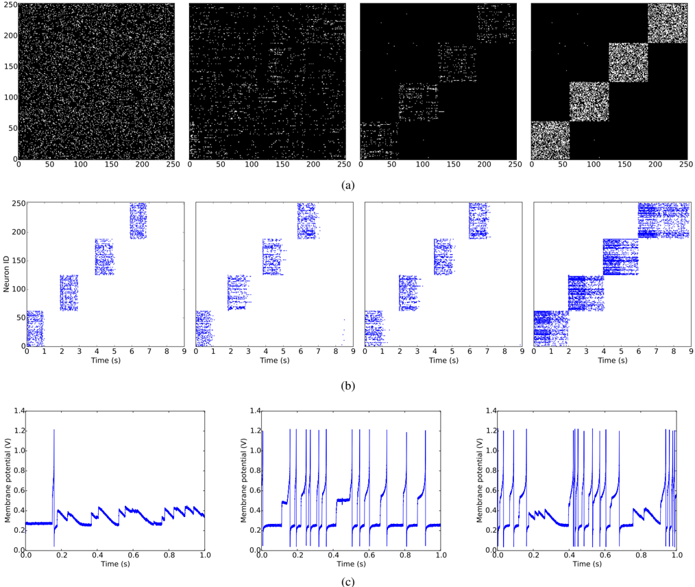
The image presents three distinct analytical sections:
1. **(a) Heatmap**: Visualizes neuronal activity distribution across time and neuron IDs.
2. **(b) Line Graphs**: Shows temporal patterns of neuronal firing rates.
3. **(c) Membrane Potential Plots**: Depicts voltage dynamics of individual neurons.
### Components/Axes
#### (a) Heatmap
- **X-axis**: Time (s) [0–250]
- **Y-axis**: Neuron ID [0–250]
- **Legend**:
- Black: No activity
- Dark gray: Low activity
- Light gray: Moderate activity
- White: High activity
- **Spatial Grounding**: Legend positioned in the top-left corner.
#### (b) Line Graphs
- **X-axis**: Time (s) [0–9]
- **Y-axis**: Neuron ID [0–250]
- **Legend**: Blue lines represent neuronal firing rates.
- **Spatial Grounding**: Legend aligned to the right of the graphs.
#### (c) Membrane Potential Plots
- **X-axis**: Time (s) [0–1]
- **Y-axis**: Membrane Potential (V) [0–1.4]
- **Legend**: Blue lines indicate voltage traces.
- **Spatial Grounding**: Legend positioned on the right.
### Detailed Analysis
#### (a) Heatmap
- **Key Trends**:
- **a1**: Uniform black (no activity) across all neurons.
- **a2**: Sparse dark gray regions (low activity) in mid-time intervals.
- **a3**: Clustered light gray regions (moderate activity) in late time intervals.
- **a4**: Structured white regions (high activity) in top-right quadrant.
- **Notable Outliers**:
- Top-left quadrant in **a4** shows reduced activity (dark gray), contrasting with the rest.
#### (b) Line Graphs
- **Key Trends**:
- **b1**: Single sharp spike at ~0.5s (Neuron ID ~50).
- **b2**: Multiple spikes at ~1s, ~3s, and ~5s (Neuron IDs ~100, ~150, ~200).
- **b3**: Gradual increase in firing rate peaking at ~7s (Neuron IDs ~50–100).
- **b4**: Dense, overlapping spikes across all time intervals (Neuron IDs ~150–250).
- **Cross-Reference**: Blue line colors match the legend; no discrepancies observed.
#### (c) Membrane Potential Plots
- **Key Trends**:
- **c1**: Single voltage spike at ~0.1s (1.2V peak).
- **c2**: Repeated spikes at ~0.2s, ~0.4s, and ~0.6s (1.0–1.1V peaks).
- **c3**: Mixed single/multiple spikes with irregular intervals (0.1–0.9s).
- **Notable Outliers**:
- **c3** shows a prolonged depolarization phase at ~0.8s, deviating from periodic patterns.
### Key Observations
1. **Heatmap-Line Graph Correlation**:
- Active regions in **a2–a4** (dark gray/white) align with spike timings in **b2–b4**.
- Example: **a4**'s top-right cluster corresponds to **b4**'s dense firing.
2. **Membrane Potential Dynamics**:
- **c1–c3** voltage spikes correlate with neuronal firing in **b1–b4**.
- **c3**'s irregular spikes suggest stochastic activity, unlike the structured patterns in **c1–c2**.
3. **Temporal Hierarchy**:
- Early time intervals (0–1s) show sparse activity (**a1**, **b1**, **c1**).
- Mid-to-late intervals (1–9s) exhibit increasing complexity (**a2–a4**, **b2–b4**, **c2–c3**).
### Interpretation
- **Neuronal Synchrony**: The heatmap (**a4**) and line graphs (**b4**) suggest synchronized firing in specific neuron clusters during late time intervals.
- **Voltage-Firing Relationship**: Membrane potential spikes (**c1–c3**) directly precede or coincide with neuronal firing events, indicating causal links between electrical activity and action potentials.
- **Anomalies**:
- **a4**'s top-left inactivity may reflect inhibitory neuron suppression.
- **c3**'s irregular spikes could indicate pathological or stress-induced neuronal behavior.
- **Implications**: The data demonstrates how temporal and spatial neuronal activity patterns emerge from underlying membrane potential dynamics, with potential applications in understanding neural coding and disorders.