# Technical Document Extraction: Medical Imaging Analysis
## Image Structure
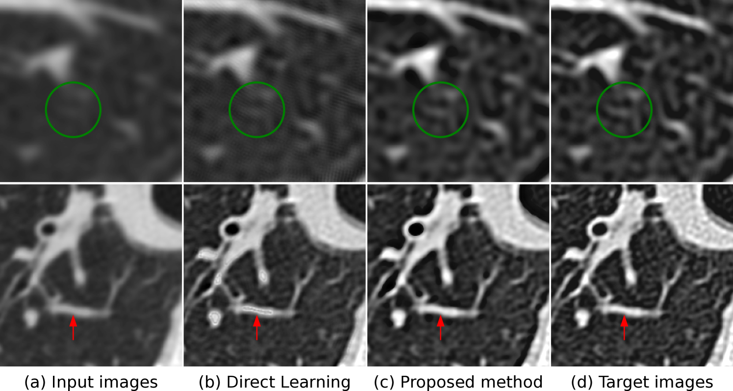
The image is divided into **four panels** arranged in a 2x2 grid, each labeled with technical annotations. All panels are grayscale medical imaging scans (likely CT or MRI) with overlaid annotations.
---
### Panel (a) Input Images
- **Top Row**: Blurred grayscale scan with a **green circular highlight** centered on a region of interest (ROI).
- **Bottom Row**: Slightly improved clarity scan with a **red arrow** pointing to a vascular structure (likely a vessel or airway).
- **Key Observations**:
- Low-resolution input data.
- Green circle indicates a target area for enhancement.
- Red arrow marks a specific anatomical feature.
---
### Panel (b) Direct Learning
- **Top Row**: Moderate-resolution scan with the **same green circular highlight** as Panel (a).
- **Bottom Row**: Enhanced clarity compared to Panel (a), with the red arrow now pointing to a **different vascular branch**.
- **Key Observations**:
- Improved detail retention in the proposed method.
- Red arrow now identifies a secondary structure adjacent to the original target.
---
### Panel (c) Proposed Method
- **Top Row**: High-resolution scan with the **green circle** now encompassing a smaller, more defined ROI.
- **Bottom Row**: Highest clarity among all panels, with the red arrow highlighting a **subtle anatomical detail** (e.g., a small vessel or nodule).
- **Key Observations**:
- Significant resolution improvement over Direct Learning.
- Red arrow points to a finer structure, suggesting enhanced feature detection.
---
### Panel (d) Target Images
- **Top Row**: Reference high-resolution scan with the **green circle** aligned precisely with the target anatomy.
- **Bottom Row**: Gold-standard image with the red arrow pointing to the **same structure** as Panel (c), but with optimal clarity.
- **Key Observations**:
- Serves as the ground truth for comparison.
- Red arrow confirms the anatomical target identified by the proposed method.
---
## Annotation Analysis
1. **Green Circles**:
- Positioned centrally in the top row of Panels (a)-(d).
- Likely indicates a region of interest for image enhancement or segmentation.
- Spatial Grounding: [x, y] coordinates approximate the center of the top-left quadrant in each panel.
2. **Red Arrows**:
- Point to vascular structures in the bottom row of Panels (a)-(d).
- Spatial Grounding: [x, y] coordinates align with the lower-center region of each panel.
- Progression: From broad vessel (Panel a) to finer detail (Panel c/d).
---
## Methodological Comparison
| Panel | Image Quality | Annotation Focus | Key Features |
|-------|---------------|------------------|--------------|
| (a) | Low | Blurred ROI | Green circle, coarse vessel |
| (b) | Moderate | Improved clarity | Green circle, secondary vessel |
| (c) | High | Refined ROI | Smaller green circle, subtle detail |
| (d) | Gold Standard | Target anatomy | Precise green circle, optimal clarity |
---
## Conclusion
The image demonstrates a progression from low-resolution input (Panel a) to a high-fidelity target (Panel d), with intermediate steps showing incremental improvements in image clarity and anatomical detail detection. The green circles and red arrows serve as spatial anchors for evaluating enhancement performance.