## Diagram: Nasal Anatomy and Surgical Markings
### Overview
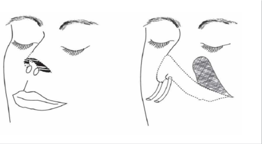
The image is a black-and-white line drawing illustrating two views of a human face, focusing on the nasal region. It appears to be a medical or surgical diagram, likely related to rhinoplasty or nasal anatomy, showing pre-operative markings or anatomical zones. There is no textual information, labels, axes, or data present in the image.
### Components/Axes
* **Left Figure:** A frontal (anterior) view of a face with closed eyes. The nose is depicted with dotted lines outlining the nasal bridge and tip. Two small, oval shapes are drawn on the nasal tip/alae region. A dark, shaded area covers the upper-left portion of the nasal dorsum (bridge).
* **Right Figure:** A three-quarter or profile (lateral) view of a face with closed eyes. The nose is outlined with a dotted line. A large, cross-hatched shaded area covers the lateral aspect of the nose, extending from the bridge down towards the cheek. Three parallel, curved lines are drawn near the base of the nose, possibly indicating incision lines or anatomical structures like the nasolabial fold.
* **Spatial Grounding:** The two figures are placed side-by-side. The frontal view is on the left, and the profile view is on the right. The shaded regions and markings are the primary informational elements.
### Detailed Analysis
* **Left Figure (Frontal View):**
* **Dotted Outline:** Traces the expected contour of the nasal bridge and tip.
* **Oval Markings:** Two small, clear ovals are positioned symmetrically on the lower nasal tip or alar rim area.
* **Shaded Region:** A dark, filled area is located on the upper-left quadrant of the nasal dorsum (from the viewer's perspective, which corresponds to the patient's right side). This likely indicates an area of focus, such as a dorsal hump to be reduced or a zone of cartilage/bone.
* **Right Figure (Profile View):**
* **Dotted Outline:** Shows the projected profile of the nose.
* **Cross-Hatched Shading:** A large, textured area covers the lateral nasal wall. This could represent a surgical flap, an area of skin undermining, or a zone of cartilage graft placement.
* **Curved Lines:** Three parallel, curved lines are drawn inferior and lateral to the nostril, following the contour of the cheek. These may represent planned incisions, skin tension lines, or the location of the nasofacial groove.
### Key Observations
1. **No Textual Data:** The diagram contains zero text—no labels, titles, legends, or annotations.
2. **Symbolic Markings:** Information is conveyed entirely through graphical symbols: dotted lines (outlines/plans), solid shading (areas of intervention), cross-hatching (a different type of tissue or procedure), and parallel lines (incisions or anatomical landmarks).
3. **Dual-Perspective Illustration:** The use of both frontal and profile views is a standard technique in surgical planning to convey three-dimensional information on a two-dimensional plane.
4. **Focus on Nasal Subunits:** The markings are concentrated on specific nasal subunits: the dorsum, tip, alae, and lateral walls.
### Interpretation
This diagram is a **surgical planning illustration for a rhinoplasty procedure**. It visually communicates the surgeon's intended modifications without the need for written text.
* **What it Demonstrates:** The markings suggest a plan that may involve:
* **Dorsal Refinement:** The shaded area on the bridge in the frontal view likely indicates a region of bone or cartilage to be shaved or reduced (a dorsal hump reduction).
* **Tip Work:** The ovals on the tip could mark points for suture placement, cartilage graft insertion, or areas of defatting.
* **Lateral Wall Modification:** The large cross-hatched area in the profile view suggests significant work on the side of the nose, possibly involving the placement of a spreader graft, a lateral crural steal technique, or the creation of a skin-soft tissue envelope flap.
* **Incision Planning:** The curved lines near the cheek may indicate the planned location for an external rhinoplasty incision or the extent of skin elevation.
* **Relationship Between Elements:** The two views are complementary. The frontal view shows symmetry and dorsal width, while the profile view shows projection, rotation, and the relationship of the nose to the cheek. The dotted lines in both likely represent the *current* anatomy, while the shaded and solid lines represent the *planned* surgical changes.
* **Anomalies/Notable Points:** The complete absence of text is notable. This implies the diagram is meant for an audience already familiar with the symbolic language of surgical planning (e.g., surgeons, medical students). The interpretation relies entirely on visual literacy within that specific field. The asymmetry of the dorsal shading in the frontal view (only on one side) is interesting and may indicate a plan to correct an asymmetric dorsal hump.