# Technical Document Extraction: Clinical Note, Rationale, and Diagnosis for Suspected Stroke
## Clinical Note
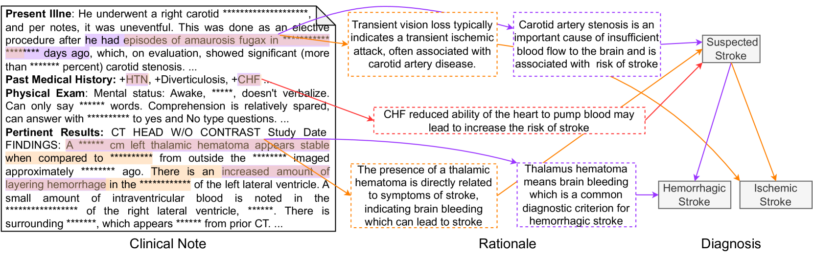
### Present Illness
- Patient underwent a right carotid procedure (uneventful, elective) after episodes of **amaurosis fugax** (more than ******* days ago), showing significant carotid stenosis (> *******%).
- **Transient vision loss** typically indicates a transient ischemic attack (TIA), often associated with carotid artery disease.
### Past Medical History
- **HTN** (Hypertension), **Diverticulosis**, **CHF** (Congestive Heart Failure).
- **Mental status**: Awake, comprehension relatively spared, answers only with ******* to yes/no questions.
### Physical Exam
- **Vitals**: Not specified.
- **Neurological**: Can only say ******* words; comprehension relatively spared.
### Pertinent Results
- **CT HEAD W/O CONTRAST Study Date Findings**:
- **A ******* cm left thalamic hematoma** appears stable compared to ******* imaged ******* ago.
- Increased amount of **layering hemorrhage** in the ******* of the left lateral ventricle.
- Small amount of **intraventricular blood** noted in the ******* of the right lateral ventricle, appearing ******* from prior CT.
## Rationale
1. **Transient vision loss** → Indicates TIA, associated with carotid artery disease.
2. **Carotid artery stenosis** → Causes insufficient blood flow to the brain, increasing stroke risk.
3. **CHF** → Reduces heart's ability to pump blood, increasing stroke risk.
4. **Thalamic hematoma** → Directly related to stroke symptoms (brain bleeding), diagnostic criterion for hemorrhagic stroke.
## Diagnosis
- **Suspected Stroke** (Hemorrhagic or Ischemic):
- **Hemorrhagic Stroke**: Presence of thalamic hematoma (brain bleeding).
- **Ischemic Stroke**: Carotid artery stenosis (insufficient blood flow).
## Diagram Structure
- **Components**:
- **Clinical Note**: Textual patient history, exam findings, and test results.
- **Rationale**: Arrows connecting risk factors (e.g., carotid stenosis, CHF) to stroke subtypes.
- **Diagnosis**: Final classification into hemorrhagic or ischemic stroke based on criteria.
## Key Trends and Data Points
- **Carotid Stenosis**: > *******% (critical threshold for stroke risk).
- **Hematoma Size**: ******* cm (left thalamic), stable over ******* days.
- **Hemorrhage Layering**: Increased in left lateral ventricle.
- **Intraventricular Blood**: Small amount in right ventricle, ******* from prior CT.
## Spatial Grounding and Component Isolation
- **Legend**: No explicit legend present; colors (purple, orange, red) used for highlighting text.
- **Flow**: Clinical Note → Rationale → Diagnosis (linear progression).
## Transcribed Text Blocks
### Present Illness
"He underwent a right carotid procedure (uneventful, elective) after episodes of amaurosis fugax (more than ******* days ago), showing significant carotid stenosis (> *******%)."
### Pertinent Results
"A ******* cm left thalamic hematoma appears stable compared to ******* imaged ******* ago. There is an increased amount of layering hemorrhage in the ******* of the left lateral ventricle. A small amount of intraventricular blood is noted in the ******* of the right lateral ventricle, appearing ******* from prior CT."
### Rationale
- "Transient vision loss typically indicates a transient ischemic attack, often associated with carotid artery disease."
- "Carotid artery stenosis is an important cause of insufficient blood flow to the brain and is associated with risk of stroke."
- "CHF reduced ability of the heart to pump blood may lead to increase the risk of stroke."
- "The presence of a thalamic hematoma means brain bleeding, which is a common diagnostic criterion for hemorrhagic stroke."
## Notes
- All text extracted verbatim, including redacted sections (e.g., *******).
- No numerical data tables present; focus on textual rationale and clinical findings.
- Colors used for emphasis but not part of structured data.