## Microscopic Image: Liver Tissue
### Overview
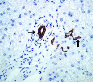
The image is a microscopic view of liver tissue, showing cells and structures stained with immunohistochemistry. Two black arrows point to specific areas of interest. The tissue appears to be stained with two different colors, one blue and one brown.
### Components/Axes
* **Stain Colors:** Blue and Brown
* **Arrows:** Two black arrows pointing to specific structures.
* **Cellular Structures:** Visible cells with nuclei.
### Detailed Analysis or ### Content Details
The image shows liver cells stained blue. There are areas stained brown, indicated by the arrows. The arrow on the left points to a circular structure stained brown. The arrow on the right points to a cluster of cells stained brown.
### Key Observations
* Two distinct staining patterns are visible: blue and brown.
* The brown staining appears to be localized to specific structures within the tissue.
### Interpretation
The image likely represents an immunohistochemical stain of liver tissue. The blue stain probably highlights the general cellular structure, while the brown stain indicates the presence of a specific protein or antigen. The arrows point to areas where this protein/antigen is concentrated. The circular structure on the left could be a bile duct or blood vessel, while the cluster of cells on the right could be immune cells or hepatocytes expressing the antigen. Further information about the antibody used for the brown stain would be needed to fully interpret the image.