\n
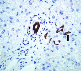
## Microscopic Image: Brain Tissue with Amyloid Plaques
### Overview
The image is a microscopic view of brain tissue, likely stained to highlight cellular structures and pathological features. The dominant feature is the presence of several amyloid plaques, indicated by darker, irregular shapes, surrounded by numerous smaller, uniformly stained cells (likely neurons and glial cells). Two plaques are specifically pointed to by black arrows. The background is a lighter color, representing the extracellular matrix.
### Components/Axes
There are no explicit axes or scales in this image. The components are:
* **Amyloid Plaques:** Irregularly shaped, dark brown/purple deposits.
* **Cells:** Small, round, blue-stained nuclei.
* **Extracellular Matrix:** Lightly stained background.
* **Arrows:** Black arrows pointing to specific amyloid plaques.
### Detailed Analysis or Content Details
The image shows a high density of cells throughout the field of view. The cells appear relatively uniform in size and staining intensity. The amyloid plaques vary in size and shape.
* **Plaque 1 (Left):** Large, roughly circular plaque with a fragmented, irregular edge. It is approximately 50-75 micrometers in diameter.
* **Plaque 2 (Center-Right):** A cluster of smaller, interconnected plaques, appearing more fragmented and less defined than the first. The combined area is approximately 30-40 micrometers.
* **Plaque 3 (Right):** A smaller, more compact plaque, approximately 15-20 micrometers in diameter, pointed to by an arrow.
* **Cell Density:** The cell density appears relatively consistent across the image, with approximately 20-30 cells visible per 100x100 micrometer square.
* **Arrow Placement:** The arrows are positioned to specifically highlight the presence of amyloid plaques.
### Key Observations
* The presence of amyloid plaques is a hallmark of Alzheimer's disease and other neurodegenerative conditions.
* The plaques are surrounded by cells, suggesting an inflammatory response.
* The variation in plaque size and shape may indicate different stages of plaque development or different types of amyloid deposits.
* The image does not provide quantitative data on plaque load or cell counts.
### Interpretation
This image likely represents brain tissue from a patient with Alzheimer's disease or a related dementia. The amyloid plaques are a key pathological feature of these conditions, and their presence suggests neuronal dysfunction and cognitive decline. The surrounding cellular response indicates an attempt by the brain to clear the plaques, but this process is often ineffective in the long term. The image demonstrates the characteristic neuropathology associated with these diseases. The lack of quantitative data limits the ability to assess the severity of the pathology, but the presence of multiple plaques suggests a significant degree of amyloid deposition. Further analysis, such as immunohistochemistry for specific proteins (e.g., beta-amyloid, tau), would be needed to confirm the diagnosis and characterize the specific type of amyloid pathology. The image is a visual confirmation of a pathological process, but does not provide information on the patient's clinical status or disease progression.