## Photograph: Histological/Cytological Slide with Stained Cellular Structures
### Overview
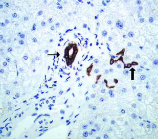
The image depicts a microscopic view of a histological or cytological sample, likely stained with hematoxylin and eosin (H&E) or similar differential staining techniques. The background is predominantly light blue, with scattered dark blue and brownish-red structures. Two black arrows highlight specific regions of interest: one pointing to a central dark blue structure and another to a cluster of brownish-red elements.
### Components/Axes
- **No explicit textual labels, axes, legends, or scale bars** are visible in the image.
- **Staining patterns**:
- **Dark blue**: Likely represents nuclei (eosinophilic staining, possibly hematoxylin).
- **Brownish-red**: May indicate cytoplasmic or extracellular matrix components (eosinophilic staining).
- **Arrows**: Two black arrows annotate specific regions but lack accompanying labels or identifiers.
### Detailed Analysis
- **Cellular morphology**:
- The dark blue structures (nuclei) vary in size and shape, with some appearing rounded and others irregular.
- The brownish-red elements are irregularly distributed, with one cluster forming a branching or dendritic pattern.
- **Staining intensity**:
- Dark blue staining is concentrated in the central region, suggesting a high density of nuclei or chromatin.
- Brownish-red staining is localized to the periphery, potentially indicating extracellular material or cytoplasmic inclusions.
- **Arrows**:
- The first arrow points to a large, dark blue structure with a clear boundary, possibly a cell nucleus or apoptotic body.
- The second arrow highlights a cluster of brownish-red elements, which may represent extracellular debris, collagen fibers, or pathological aggregates.
### Key Observations
1. **Lack of scale**: No scale bar is present, making it impossible to quantify cell size or spacing.
2. **Staining ambiguity**: Without a legend, the exact identity of the stains (e.g., hematoxylin, eosin, or other markers) cannot be confirmed.
3. **Pathological features**: The irregular clustering of brownish-red elements and the prominence of dark blue nuclei may suggest pathological conditions (e.g., inflammation, fibrosis, or malignancy), but this requires contextual clinical or experimental data.
### Interpretation
- **Biological significance**:
- The dark blue nuclei likely represent viable cells, while the brownish-red elements could indicate extracellular matrix deposition, cellular debris, or apoptotic material.
- The arrows may highlight areas of interest for further analysis (e.g., tumor cell clusters, inflammatory infiltrates, or tissue damage).
- **Limitations**:
- The absence of textual annotations, legends, or scale bars limits the ability to draw definitive conclusions.
- Differential staining patterns alone cannot confirm specific cell types or pathologies without additional context (e.g., immunohistochemistry markers, clinical history).
- **Next steps**:
- Cross-reference with experimental protocols to identify staining agents and their targets.
- Compare with control samples to assess pathological changes.
**Note**: This image contains no explicit textual data, numerical values, or structured information. The analysis is based solely on visual interpretation of staining patterns and morphological features.