# Technical Document Extraction: MoSe2 Fermi Energy Heatmaps
## Image Description
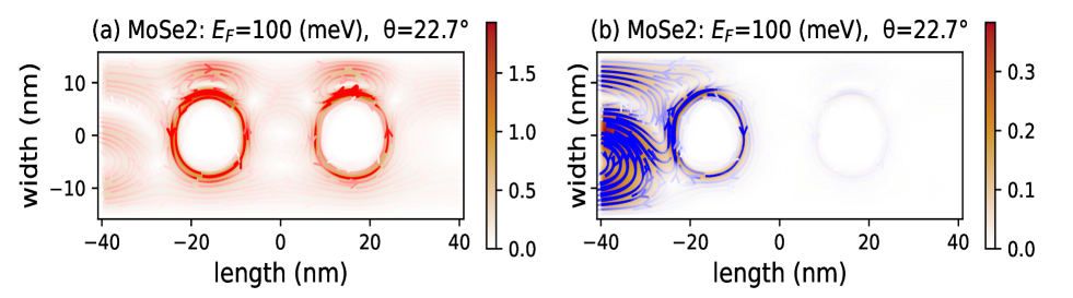
The image contains two side-by-side heatmaps labeled **(a)** and **(b)**, depicting Fermi energy distributions in MoSe2 at a fixed angle **θ = 22.7°**. Both panels share identical axis labels and spatial dimensions but differ in Fermi energy values (**E_F**) and intensity scales.
---
### **Panel (a): E_F = 100 meV**
- **Title**: `MoSe2: E_F=100 (meV), θ=22.7°`
- **Axis Labels**:
- **X-axis**: `length (nm)` (range: -40 to 40 nm)
- **Y-axis**: `width (nm)` (range: -10 to 10 nm)
- **Color Scale**:
- **Legend**: Red-to-white gradient (0.0 to 1.5)
- **Key Observation**: Two bright red circular regions centered at approximately (±20 nm, 0 nm) with high-intensity gradients radiating outward.
- **Arrows**: Red directional vectors indicating flow or gradient direction, concentrated near the circular regions.
---
### **Panel (b): E_F = 100 meV**
- **Title**: `MoSe2: E_F=100 (meV), θ=22.7°`
- **Axis Labels**:
- **X-axis**: `length (nm)` (range: -40 to 40 nm)
- **Y-axis**: `width (nm)` (range: -10 to 10 nm)
- **Color Scale**:
- **Legend**: Blue-to-white gradient (0.0 to 0.3)
- **Key Observation**: A single faint blue circular region centered at approximately (0 nm, 0 nm) with weaker intensity and broader spatial distribution compared to Panel (a).
- **Arrows**: Blue directional vectors, less dense and more dispersed than in Panel (a).
---
### **Cross-Referenced Observations**
1. **Legend Consistency**:
- Panel (a) uses **red** to denote higher intensity (max 1.5), while Panel (b) uses **blue** for lower intensity (max 0.3).
- Arrows in both panels match their respective color schemes (red in (a), blue in (b)).
2. **Spatial Trends**:
- Panel (a) shows localized, high-intensity features, suggesting strong Fermi surface localization.
- Panel (b) exhibits diffuse intensity, indicating broader Fermi surface dispersion.
3. **Angle Consistency**: Both panels share the same **θ = 22.7°**, implying comparative analysis under identical crystallographic orientations.
---
### **Summary**
The heatmaps contrast Fermi energy distributions in MoSe2 at **E_F = 100 meV** under identical angular conditions. Panel (a) highlights localized, high-intensity regions, while Panel (b) demonstrates broader, lower-intensity distributions. The color scales and directional arrows provide quantitative and qualitative insights into electronic behavior.