## Medical Diagnosis Procedure Flowchart
### Overview
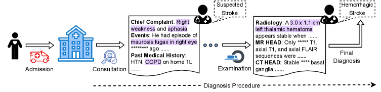
This image is a technical flowchart illustrating a medical diagnostic pathway for a patient presenting with stroke symptoms. It depicts a linear process from hospital admission to final diagnosis, incorporating clinical information, examination steps, and radiological findings. The diagram uses icons, text boxes, and directional arrows to map the sequence of events and data points.
### Components/Axes
The flowchart is organized horizontally from left to right, representing chronological progression. Key components are:
1. **Process Start (Left):**
* **Icons:** A person silhouette, an ambulance, and a hospital building.
* **Label:** "Admission" (below the icons).
* **Arrow:** Points right to the next stage.
2. **Consultation Stage:**
* **Icon:** A magnifying glass over a document.
* **Label:** "Consultation" (below the icon).
* **Arrow:** Points right to the main clinical data box.
3. **Clinical Data Box (Center-Left):**
* **Title:** "Chief Complaint: Right weakness and aphasia."
* **Sub-sections:**
* "Events: He had episode of amaurosis fugax in right eye ......... ago"
* "Past Medical History: HTN, COPD on home 1L"
* **Icon:** A stethoscope icon is placed to the left of this box.
4. **Examination Stage:**
* **Icon:** A computer monitor with a magnifying glass.
* **Label:** "Examination" (below the icon).
* **Arrow:** Points right to the radiology findings box.
5. **Radiology Findings Box (Center-Right):**
* **Title:** "Radiology: A 3.0 x 1.1 cm left thalamic hematoma appears stable when ........."
* **Sub-sections:**
* "MR HEAD: Only **** T1, axial T1, and axial FLAIR sequences were ........."
* "CT HEAD: Stable *** basal ganglia ........."
* **Icon:** A stethoscope icon is placed to the left of this box.
6. **Diagnostic Icons & Final Output (Right):**
* **Top Icon:** A brain with a lightning bolt, labeled "Suspected Stroke" in a dashed box above it.
* **Bottom Icon:** A brain with a red spot, labeled "Hemorrhagic Stroke" in a dashed box above it.
* **Final Label:** "Final Diagnosis" (below the hemorrhagic stroke icon).
* **Arrows:** A dashed arrow connects the "Suspected Stroke" icon to the "Hemorrhagic Stroke" icon. A solid arrow points from the radiology box to the "Final Diagnosis" label.
7. **Overall Process Label:**
* A long, dashed arrow runs along the bottom of the entire diagram, labeled "Diagnosis Procedure".
### Detailed Analysis / Content Details
**Transcribed Text with Uncertainty:**
* **Chief Complaint Box:**
* "Chief Complaint: Right weakness and aphasia." (Clear)
* "Events: He had episode of amaurosis fugax in right eye ......... ago" (The duration is obscured by dots).
* "Past Medical History: HTN, COPD on home 1L" (HTN = Hypertension, COPD = Chronic Obstructive Pulmonary Disease, 1L = likely 1 Liter of supplemental oxygen).
* **Radiology Box:**
* "Radiology: A 3.0 x 1.1 cm left thalamic hematoma appears stable when ........." (The comparison timeframe is obscured).
* "MR HEAD: Only **** T1, axial T1, and axial FLAIR sequences were ........." (The number of sequences is obscured by asterisks; the conclusion is incomplete).
* "CT HEAD: Stable *** basal ganglia ........." (The specific finding or region is partially obscured).
**Spatial Grounding & Flow:**
* The legend (labels for "Suspected Stroke" and "Hemorrhagic Stroke") is positioned in the top-right quadrant, directly above their corresponding brain icons.
* The flow is strictly linear: Admission -> Consultation -> Clinical Data -> Examination -> Radiology Data -> Final Diagnosis.
* The "Suspected Stroke" icon is placed above and before the "Hemorrhagic Stroke" icon, connected by a dashed arrow, indicating a diagnostic refinement from a general suspicion to a specific type.
### Key Observations
1. **Diagnostic Refinement:** The pathway shows a clear progression from a broad "Suspected Stroke" to a specific "Hemorrhagic Stroke" diagnosis, driven by the radiological finding of a "left thalamic hematoma."
2. **Critical Data Points:** The key quantitative data is the size of the hematoma: **3.0 x 1.1 cm**. Its location is specified as the **left thalamus**.
3. **Clinical Context:** The patient's history (HTN, COPD) and presenting symptoms (right-sided weakness, aphasia, prior episode of amaurosis fugax) provide essential context for the stroke workup.
4. **Incomplete Information:** Several text fields are intentionally obscured with dots or asterisks, indicating that specific details (timeframes, exact sequence counts, precise CT findings) are either redacted or meant to be filled in.
### Interpretation
This flowchart is a schematic representation of a standard acute stroke diagnostic protocol. It demonstrates how clinical history (past medical conditions, symptom onset) and physical examination findings are integrated with advanced neuroimaging (MRI and CT) to arrive at a definitive diagnosis.
The data suggests a patient with vascular risk factors (hypertension) presenting with acute neurological deficits. The imaging confirms a hemorrhagic stroke (bleeding) in a deep brain structure (thalamus), which explains the contralateral (right-sided) weakness. The notation that the hematoma "appears stable" is crucial, as it informs immediate management decisions, such as blood pressure control and the need for neurosurgical consultation.
The diagram's structure emphasizes a methodical, evidence-based approach: subjective patient data leads to objective imaging, which in turn confirms the initial clinical suspicion and specifies the pathology. The obscured text highlights that while the core diagnostic facts (size, location, type) are clear, ancillary details are part of a larger, more detailed medical record.