## Diagram: Stroke Diagnosis Flowchart
### Overview
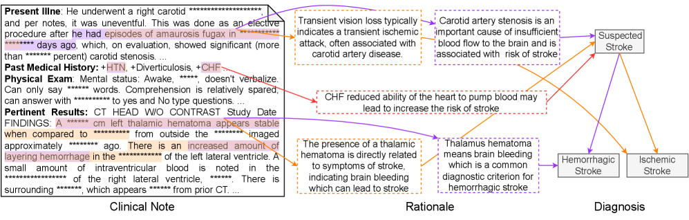
This diagram illustrates the diagnostic pathway from clinical notes and findings to a suspected stroke diagnosis, differentiating between hemorrhagic and ischemic stroke. It connects clinical observations (left), the rationale behind the diagnosis (center), and the resulting diagnosis (right). The diagram uses a flow-chart style with arrows indicating the relationships between observations, reasoning, and diagnoses. Significant portions of the text are obscured with asterisks ("****").
### Components/Axes
The diagram is divided into three main sections:
* **Clinical Note (Left):** Contains patient history and examination findings.
* **Rationale (Center):** Explains the reasoning connecting clinical findings to diagnoses.
* **Diagnosis (Right):** Presents the possible diagnoses: Suspected Stroke, Hemorrhagic Stroke, and Ischemic Stroke.
The diagram uses colored arrows to represent the flow of information:
* **Blue:** Connects clinical notes to suspected stroke and ischemic stroke.
* **Red:** Connects clinical notes to suspected stroke and hemorrhagic stroke.
* **Yellow:** Connects clinical notes to rationale.
* **Green:** Connects rationale to hemorrhagic stroke.
### Detailed Analysis or Content Details
**Clinical Note:**
* "Present Illne: He underwent a right carotid **** and per notes, it was uneventful. This was done as an elective procedure after he had episodes of amaurosis fugax in **** days ago, which, on evaluation, showed significant (more than **** percent) carotid stenosis..."
* "Past Medical History: +HTN, +Diverticulosis, +CHF..."
* "Physical Exam Mental status: Awake, **** doesn't verbalize. Can only say **** words. Comprehension is relatively spared, can answer with **** to yes and no type questions..."
* "Pertinent Results: CT HEAD W/O CONTRAST Study Date FINDINGS. A **** cm left thalamic hematoma appears stable when compared to **** from outside the **** imaged approximately **** ago. There is an increased amount of layering hemorrhage in the **** of the left lateral ventricle. A small amount of intraventricular blood is noted in the **** of the right lateral ventricle, ****. There is surrounding ****, which appears **** from prior CT..."
**Rationale:**
* "Transient vision loss typically indicates a transient ischemic attack, often associated with carotid artery disease." (Connected to Suspected Stroke and Ischemic Stroke via blue arrow)
* "Carotid artery stenosis is an important cause of insufficient blood flow to the brain and is associated with stroke." (Connected to Suspected Stroke and Ischemic Stroke via blue arrow)
* "CHF reduced ability of the heart to pump blood may lead to increase the risk of stroke." (Connected to Suspected Stroke and Hemorrhagic Stroke via red arrow)
* "The presence of a thalamic hematoma is directly related to symptoms of stroke, indicating brain bleeding which can lead to stroke." (Connected to Hemorrhagic Stroke via green arrow)
* "Thalamus hematoma means brain bleeding which is a common diagnostic criterion for hemorrhagic stroke." (Connected to Hemorrhagic Stroke via green arrow)
**Diagnosis:**
* Suspected Stroke (Connected to Clinical Note via blue and red arrows)
* Hemorrhagic Stroke (Connected to Clinical Note via red arrow and Rationale via green arrow)
* Ischemic Stroke (Connected to Clinical Note via blue arrow)
### Key Observations
* The diagram highlights multiple pathways leading to a stroke diagnosis.
* The presence of a thalamic hematoma strongly suggests hemorrhagic stroke.
* Carotid stenosis and CHF are risk factors associated with both ischemic and hemorrhagic stroke.
* Significant portions of the clinical note are redacted, limiting the completeness of the information.
* The diagram suggests that a suspected stroke diagnosis can lead to either ischemic or hemorrhagic stroke, depending on the clinical findings and rationale.
### Interpretation
The diagram represents a simplified clinical decision-making process for stroke diagnosis. It demonstrates how clinical observations, such as carotid stenosis, CHF, and the presence of a thalamic hematoma, are used to formulate a rationale for a diagnosis. The diagram emphasizes the importance of differentiating between ischemic and hemorrhagic stroke, as the treatment strategies differ significantly. The obscured text suggests that the full clinical picture is more complex than what is presented in the diagram. The diagram serves as a visual aid for understanding the relationships between clinical findings, reasoning, and diagnoses in stroke patients. The multiple pathways indicate that stroke diagnosis is not always straightforward and requires careful consideration of all available information. The diagram is a high-level overview and does not include details about the specific diagnostic tests or criteria used to confirm a stroke diagnosis.