## Medical Flowchart: Acute Coronary Syndrome (ACS) Diagnostic Pathway
### Overview
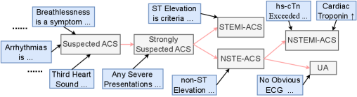
The image is a black-and-white flowchart diagram illustrating a clinical decision-making pathway for diagnosing Acute Coronary Syndrome (ACS). It maps the progression from initial symptoms and presentations to specific diagnostic categories (STEMI-ACS, NSTEMI-ACS, UA). The diagram uses rectangular boxes for concepts/criteria and directional arrows to indicate logical flow and relationships.
### Components/Axes
The diagram is structured horizontally, flowing generally from left (initial presentation) to right (final diagnosis). It can be segmented into three logical regions:
1. **Left Region (Initial Presentation & Suspicion):** Contains boxes listing symptoms and initial assessments.
2. **Middle Region (Diagnostic Criteria & Strong Suspicion):** Contains boxes for key diagnostic criteria and the "Strongly Suspected ACS" node.
3. **Right Region (Final ACS Classification):** Contains the final diagnostic categories.
**All Textual Elements (Transcribed):**
* `Breathlessness is a symptom`
* `Arrhythmias is ....`
* `Third Heart Sound ...`
* `Suspected ACS`
* `Any Severe Presentations ...`
* `ST Elevation is criteria ...`
* `Strongly Suspected ACS`
* `non-ST Elevation ...`
* `STEMI-ACS`
* `NSTE-ACS`
* `hs-cTnT Exceeded ...`
* `Cardiac Troponin ↑`
* `NSTEMI-ACS`
* `No Obvious ECG ...`
* `UA`
**Flow Connections (Arrows):**
* `Breathlessness is a symptom` → `Suspected ACS`
* `Arrhythmias is ....` → `Suspected ACS`
* `Third Heart Sound ...` → `Suspected ACS`
* `Suspected ACS` → `Strongly Suspected ACS`
* `Any Severe Presentations ...` → `Strongly Suspected ACS`
* `ST Elevation is criteria ...` → `Strongly Suspected ACS`
* `ST Elevation is criteria ...` → `STEMI-ACS`
* `Strongly Suspected ACS` → `STEMI-ACS`
* `Strongly Suspected ACS` → `NSTE-ACS`
* `non-ST Elevation ...` → `NSTE-ACS`
* `NSTE-ACS` → `NSTEMI-ACS` (via path involving `hs-cTnT Exceeded ...` and `Cardiac Troponin ↑`)
* `NSTE-ACS` → `UA` (via path involving `No Obvious ECG ...`)
### Detailed Analysis
The diagram outlines a stepwise diagnostic logic:
1. **Initial Suspicion (`Suspected ACS`):** This is triggered by the presence of non-specific symptoms like breathlessness, arrhythmias, or a third heart sound.
2. **Escalation to Strong Suspicion (`Strongly Suspected ACS`):** Suspicion is heightened by "Any Severe Presentations" or the specific criterion of "ST Elevation."
3. **Branching to Specific Diagnoses:**
* **STEMI-ACS (ST-Elevation Myocardial Infarction):** Directly linked to the "ST Elevation is criteria" box and also stemming from "Strongly Suspected ACS."
* **NSTE-ACS (Non-ST-Elevation ACS):** This is a broader category stemming from "Strongly Suspected ACS" and also linked to "non-ST Elevation." It then bifurcates based on biomarker and ECG findings:
* **NSTEMI-ACS (Non-ST-Elevation Myocardial Infarction):** The path involves the boxes `hs-cTnT Exceeded ...` and `Cardiac Troponin ↑`, indicating elevated cardiac troponin levels.
* **UA (Unstable Angina):** The path involves the box `No Obvious ECG ...`, suggesting the absence of diagnostic ECG changes or significant troponin elevation.
### Key Observations
* **Ellipses (...):** Multiple boxes end with ellipses (e.g., `Arrhythmias is ....`, `hs-cTnT Exceeded ...`). This indicates the text is truncated or represents a placeholder for a more detailed list or explanation not fully shown in this diagram.
* **Hierarchical Structure:** The flowchart shows a clear hierarchy: Symptoms → Suspicion → Strong Suspicion → Specific Diagnosis.
* **Critical Decision Points:** "ST Elevation" is a pivotal criterion that directly leads to a STEMI diagnosis and also contributes to "Strongly Suspected ACS." The differentiation between NSTEMI and UA hinges on the presence of elevated cardiac troponins (`Cardiac Troponin ↑`) versus their absence or lack of obvious ECG changes.
* **Visual Layout:** The "Strongly Suspected ACS" box acts as a central hub, connecting the initial suspicion phase to the final diagnostic categories. The final diagnoses (STEMI-ACS, NSTEMI-ACS, UA) are positioned on the far right.
### Interpretation
This diagram represents a simplified clinical algorithm for the triage and diagnosis of Acute Coronary Syndrome. It visually encodes the standard medical approach where:
1. Non-specific symptoms raise initial suspicion.
2. Specific high-risk features (severe presentations, ST-elevation) significantly increase diagnostic certainty.
3. The final classification depends on a combination of **ECG findings** (ST-elevation vs. non-ST-elevation/no obvious changes) and **cardiac biomarker results** (troponin elevation).
The pathway emphasizes that **STEMI** is diagnosed primarily on ECG criteria (ST-elevation), allowing for rapid intervention. **NSTEMI** and **UA** are both under the NSTE-ACS umbrella but are distinguished by the presence of myocardial necrosis, as evidenced by elevated troponins in NSTEMI. The ellipses suggest this is a high-level overview, and a complete clinical protocol would include more detailed criteria for each box (e.g., specific troponin thresholds, definitions of "severe presentations"). The diagram's purpose is to map the logical relationships between clinical findings and diagnostic labels, serving as a conceptual guide for healthcare professionals.