\n
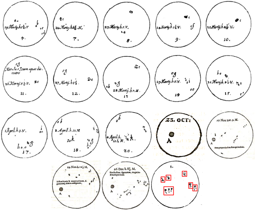
## Petri Dish Observations: Bacterial Growth Study
### Overview
The image depicts a series of 21 petri dishes, each containing bacterial growth. Each dish is labeled with a date, initials, and potentially strain information. The dishes appear to document a time-series experiment observing bacterial colony formation. The bottom row contains a key to symbols used within the dishes.
### Components/Axes
The image lacks traditional axes. The "axes" are the arrangement of the petri dishes in a roughly 3x7 grid. Each dish is labeled with the following information:
* **Number:** 1-21, sequentially arranged.
* **Date:** Appears in the format "Month. Day" (e.g., "23. OCT").
* **Initials:** A combination of letters (e.g., "h.s.v.", "h.M.").
* **Strain/Description:** Additional text describing the bacterial strain or observation (e.g., "Clavula r. Imum aper. dis.", "Diphtheriae & squamosae...").
* **Symbols:** Small symbols within the dishes represent specific observations.
The bottom row (dishes 19-21) contains a key explaining the symbols:
* **A:** A square with a diagonal line.
* **B:** A square with a cross.
* **C:** A square with a dot.
* **D:** A square with a diagonal line and a dot.
### Detailed Analysis or Content Details
Here's a transcription of the labels for each petri dish:
1. "19. März h.s.v. i"
2. "20. März h.s.v. i"
3. "21. März h.s.v. i"
4. "22. März h.s.v. i"
5. "23. März h.s.v. i"
6. "Clavula r. Imum aper. dis."
7. "26. März h.s.v. &"
8. "27. März h.s.v."
9. "28. März h.s.v. i"
10. "29. März h.s.v. i"
11. "30. März h.s.v. &"
12. "April. h.s.v. i"
13. "2. April. h.s.v."
14. "3. April. h.s.v."
15. "4. April. h.M. &"
16. "17. Nou. h.s.v. i"
17. "18. Nou. h.s.v. i"
18. "19. Nou. h.s.v. i"
19. "20. Dec. h.s.v. M. Diphtheriae & squamosae..."
20. "21. Dec. h.s.v. M. Bacilla from specimen, malignant..."
21. "23. OCT: A B C D"
The dishes show varying degrees of bacterial growth, ranging from sparse colonies to more confluent growth. The symbols within the dishes (A, B, C, D) appear to indicate different characteristics of the colonies, such as morphology or staining properties.
### Key Observations
* The experiment appears to have started in March and continued into December, with a gap in October.
* The "h.s.v." strain appears to be the most frequently studied, appearing in multiple dishes across different dates.
* The dishes labeled "April" and "December" show different growth patterns and potentially different strains.
* The key in the bottom row suggests a systematic way of categorizing the observed colonies.
### Interpretation
This image documents a microbiological study, likely focused on identifying and characterizing bacterial strains. The sequential labeling of the petri dishes suggests a time-course experiment, where the growth of bacteria was monitored over several months. The use of initials ("h.s.v.", "h.M.") likely refers to the researcher or the source of the bacterial cultures. The symbols (A, B, C, D) represent a qualitative assessment of the colonies, potentially based on their appearance under a microscope.
The experiment seems to have involved multiple strains, including "Clavula r. Imum aper. dis." and strains isolated from specimens ("Diphtheriae & squamosae...", "Bacilla from specimen, malignant..."). The varying growth patterns observed in the dishes suggest differences in the growth rates or environmental requirements of the different strains. The gap in October might represent a pause in the experiment or a change in the experimental protocol.
The image provides a snapshot of a scientific investigation, highlighting the importance of careful observation and documentation in microbiological research. The data suggests a systematic approach to studying bacterial growth and characterization, using both quantitative (time-series data) and qualitative (symbol-based assessment) methods. The image is a historical record of early microbiological work, demonstrating the methods used to study bacteria before the advent of modern techniques.