## Biological Neuron Diagram: Neuronal Structure and Synaptic Transmission
### Overview
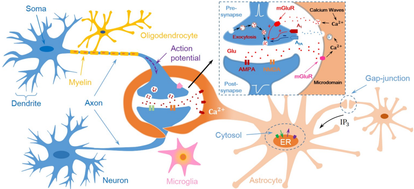
The image is a detailed biological diagram illustrating the structure of a neuron, its components, and the process of synaptic transmission. It includes labeled parts of a neuron (e.g., soma, dendrite, axon), glial cells (oligodendrocyte, microglia, astrocyte), and a zoomed-in inset detailing synaptic mechanisms. Colors are used to differentiate cell types and processes (e.g., blue for neuron structures, yellow for oligodendrocytes, pink for microglia).
---
### Components/Axes
#### Labels and Elements:
1. **Neuron Components**:
- **Soma**: Cell body of the neuron.
- **Dendrite**: Branched extensions receiving signals.
- **Axon**: Long projection transmitting signals.
- **Myelin**: Insulating sheath around the axon (produced by oligodendrocytes).
- **Axon Terminals**: Endpoints releasing neurotransmitters.
- **Oligodendrocyte**: Glial cell myelinating axons (yellow).
- **Microglia**: Immune cells (pink star-shaped).
- **Astrocyte**: Star-shaped glial cell supporting synapses (orange).
2. **Synaptic Transmission Inset**:
- **Pre-synapse**: Contains vesicles with neurotransmitters (e.g., glutamate, GABA).
- **Post-synapse**: Includes ion channels (AMPA, NMDA receptors) and calcium waves.
- **Calcium Waves**: Triggered by IP₃ signaling, leading to exocytosis.
- **Gap-junction**: Direct communication between astrocytes (orange).
3. **Key Processes**:
- **Action Potential**: Depolarization wave along the axon.
- **Exocytosis**: Release of neurotransmitters (e.g., glutamate) into the synaptic cleft.
- **Ion Channels**: mGluR (metabotropic glutamate receptor), AMPA, NMDA (ionotropic receptors).
- **Second Messenger System**: IP₃ (inositol trisphosphate) and Ca²⁺ (calcium ions) signaling.
---
### Detailed Analysis
#### Neuron Structure:
- **Soma**: Central hub for integrating signals.
- **Dendrites**: Receive synaptic inputs; branched structure increases surface area.
- **Axon**: Conducts electrical impulses away from the soma. Myelinated axons (insulated by oligodendrocytes) enable faster signal propagation.
- **Axon Terminals**: Release neurotransmitters (e.g., glutamate) into the synaptic cleft via exocytosis.
#### Synaptic Transmission (Inset):
1. **Pre-synaptic Events**:
- Action potential arrives at the axon terminal.
- Voltage-gated Ca²⁺ channels open, allowing Ca²⁺ influx.
- Ca²⁺ binds to sensors, triggering vesicle fusion with the membrane (exocytosis).
- Neurotransmitters (e.g., glutamate) are released into the synaptic cleft.
2. **Post-synaptic Events**:
- Neurotransmitters bind to receptors (AMPA, NMDA, mGluR).
- **AMPA Receptors**: Allow Na⁺ influx, depolarizing the post-synaptic neuron.
- **NMDA Receptors**: Require both glutamate and post-synaptic depolarization to open, permitting Ca²⁺ influx.
- **mGluR**: G-protein-coupled receptors initiating second messenger pathways (e.g., IP₃ production).
- **Calcium Waves**: IP₃ triggers Ca²⁺ release from intracellular stores, amplifying signaling.
3. **Glial Cell Roles**:
- **Oligodendrocytes**: Myelinate axons, increasing conduction speed.
- **Astrocytes**: Maintain synaptic homeostasis via gap-junctional communication and uptake of neurotransmitters.
- **Microglia**: Monitor neuronal health and respond to damage/inflammation.
---
### Key Observations
1. **Signal Propagation**: The neuron’s structure (dendrites → axon → terminals) ensures unidirectional signal flow.
2. **Synaptic Complexity**: Multiple receptor types (AMPA, NMDA, mGluR) allow diverse post-synaptic responses.
3. **Glial Integration**: Astrocytes and oligodendrocytes are critical for synaptic function and myelination.
4. **Calcium Dynamics**: Ca²⁺ acts as a key second messenger, linking extracellular signals to intracellular responses.
---
### Interpretation
This diagram highlights the neuron as a highly specialized cell for electrical and chemical signaling. The interplay between neurons and glial cells (oligodendrocytes, astrocytes, microglia) underscores the complexity of neural networks. The synaptic transmission process, involving Ca²⁺-dependent exocytosis and receptor-mediated signaling, demonstrates how neurons integrate and propagate information. The inset’s focus on ion channels and second messengers (e.g., IP₃) emphasizes the molecular precision required for synaptic plasticity and learning. Notably, the absence of numerical data suggests this is a conceptual model rather than a quantitative analysis, prioritizing structural and functional relationships over measurable metrics.