## Clinical Decision Pathway: Acute Coronary Syndrome (ACS) Diagnosis
### Overview
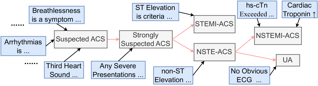
The image is a clinical flowchart or decision pathway diagram illustrating the diagnostic process for Acute Coronary Syndrome (ACS). It maps the progression from initial symptoms and clinical findings to specific ACS diagnoses, using a combination of blue boxes (representing symptoms, signs, or criteria) and gray boxes (representing diagnostic states or categories). The flow moves generally from left to right, with arrows indicating the logical progression and branching based on specific criteria.
### Components/Axes
The diagram is structured into three main conceptual regions:
1. **Left Region (Initial Presentation):** Contains blue boxes listing symptoms and signs that lead to a suspicion of ACS.
2. **Central Region (Diagnostic Progression):** Contains gray boxes representing escalating levels of diagnostic certainty ("Suspected ACS" -> "Strongly Suspected ACS") and the primary branching point for ACS types.
3. **Right Region (Final Classifications):** Contains gray boxes for the final diagnostic categories (STEMI-ACS, NSTE-ACS, NSTEMI-ACS, UA) and blue boxes listing the specific criteria that lead to each.
**Legend/Color Coding:**
* **Blue Boxes:** Clinical symptoms, signs, or diagnostic criteria (e.g., "Breathlessness is a symptom...", "ST Elevation is criteria...").
* **Gray Boxes:** Diagnostic states or categories (e.g., "Suspected ACS", "STEMI-ACS").
* **Arrows:** Indicate the flow of clinical reasoning. Black arrows show contributing factors or criteria leading to a state. Red arrows show the primary diagnostic pathway progression.
### Detailed Analysis
**Textual Content & Flow:**
**1. Initial Symptoms & Signs (Left Region - Blue Boxes):**
* "Breathlessness is a symptom ..." (Top-left, points to "Suspected ACS")
* "Arrhythmias is ..." (Far-left, points to "Suspected ACS")
* "Third Heart Sound ..." (Bottom-left, points to "Suspected ACS")
* "Any Severe Presentations ..." (Bottom-center, points to "Strongly Suspected ACS")
**2. Diagnostic States (Gray Boxes - Central Flow):**
* **Suspected ACS:** The initial diagnostic state, reached from the symptoms listed above.
* **Strongly Suspected ACS:** The next state, reached from "Suspected ACS" (red arrow) and informed by "Any Severe Presentations..." (black arrow).
**3. Branching Criteria & Final Diagnoses (Right Region):**
From "Strongly Suspected ACS," the pathway branches via red arrows:
* **Branch 1 (Top Path):**
* **Criteria (Blue Box):** "ST Elevation is criteria ..." (Points to "STEMI-ACS")
* **Diagnosis (Gray Box):** **STEMI-ACS** (ST-Elevation Myocardial Infarction - Acute Coronary Syndrome)
* **Branch 2 (Bottom Path):**
* **Criteria (Blue Box):** "non-ST Elevation ..." (Points to "NSTE-ACS")
* **Diagnosis (Gray Box):** **NSTE-ACS** (Non-ST-Elevation Acute Coronary Syndrome)
From **NSTE-ACS**, the pathway further differentiates:
* **Sub-Branch 2a (Top Path from NSTE-ACS):**
* **Criteria (Blue Boxes):**
* "hs-cTn Exceeded ..." (High-sensitivity cardiac Troponin)
* "Cardiac Troponin ↑" (Increased)
* **Diagnosis (Gray Box):** **NSTEMI-ACS** (Non-ST-Elevation Myocardial Infarction - ACS)
* **Sub-Branch 2b (Bottom Path from NSTE-ACS):**
* **Criteria (Blue Box):** "No Obvious ECG ..." (Electrocardiogram)
* **Diagnosis (Gray Box):** **UA** (Unstable Angina)
### Key Observations
1. **Hierarchical Diagnosis:** The flowchart presents a clear hierarchy: from general suspicion ("Suspected ACS") to strong suspicion, and then to specific, mutually exclusive final diagnoses (STEMI, NSTEMI, UA).
2. **Critical Decision Points:** The two major branching points are:
* The presence or absence of **ST Elevation** on ECG, which separates STEMI from NSTE-ACS.
* Within NSTE-ACS, the status of **cardiac troponin** levels, which separates NSTEMI (elevated troponin) from Unstable Angina (no obvious ECG changes, implying troponin is not elevated or the presentation is primarily ischemic without biomarker rise).
3. **Symptom vs. Criteria:** The initial blue boxes list non-specific symptoms (breathlessness, arrhythmias, third heart sound) that raise suspicion. The later blue boxes list more specific diagnostic criteria (ST elevation, troponin levels) that confirm a specific diagnosis.
4. **Incomplete Text:** Several blue boxes contain ellipses ("..."), indicating that the text is truncated. The full criteria or descriptions are not visible in this diagram.
### Interpretation
This flowchart represents a standardized clinical algorithm for triaging and diagnosing patients with suspected Acute Coronary Syndrome. It visually encodes the **Peircean investigative logic** of medical diagnosis:
* **Abduction:** Initial symptoms (breathlessness, arrhythmias) lead to the abductive inference of a possible ACS ("Suspected ACS").
* **Deduction:** Based on the established diagnostic rules (e.g., "If ST elevation is present, then it is STEMI"), the clinician deduces the specific type of ACS from the "Strongly Suspected" state.
* **Induction:** The final diagnosis (e.g., NSTEMI) is confirmed by specific, observable facts (elevated hs-cTn), which inductively support the general category.
The diagram emphasizes that ACS is not a single entity but a spectrum. The critical differentiation between **STEMI** (a full-thickness heart attack requiring immediate reperfusion) and **NSTE-ACS** (which includes NSTEMI and UA) is driven primarily by the ECG finding of ST elevation. The subsequent split between **NSTEMI** and **UA** hinges on myocardial necrosis, as evidenced by elevated cardiac troponins. The pathway underscores the importance of integrating clinical presentation, ECG findings, and biomarker results to arrive at an accurate diagnosis, which is essential for determining the correct and urgent treatment strategy. The truncated text suggests this is a simplified overview, and the full criteria would be detailed in accompanying medical guidelines.