\n
## Micrograph: Nanostructured Material
### Overview
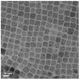
The image is a grayscale micrograph depicting a highly ordered, two-dimensional nanostructure. The structure consists of a repeating pattern of interconnected square or rectangular cells, resembling a lattice or mesh. The image appears to be a transmission electron microscopy (TEM) image, given the high contrast and resolution.
### Components/Axes
The only explicit label present is a scale bar located in the bottom-left corner, indicating a length of "10 nm". There are no axes or legends present. The image itself represents the sample being observed.
### Detailed Analysis or Content Details
The nanostructure is composed of dark lines forming the walls of the cells, and lighter areas within the cells. The cells are approximately square, with an estimated side length of 2-3 nm. The lines appear to have a consistent width throughout the image. The structure is highly uniform, with minimal defects or irregularities visible. The overall image is approximately 500x500 pixels. The contrast between the cell walls and the interior is high, suggesting a difference in material composition or density.
### Key Observations
The most striking observation is the high degree of order and uniformity in the nanostructure. The repeating pattern suggests a self-assembly process or a highly controlled fabrication method. The scale bar indicates that the features are in the nanometer range, confirming the nanoscale nature of the structure. There are no obvious variations in cell size or shape across the image.
### Interpretation
The image likely represents a synthesized material with a designed nanostructure. The regular arrangement of cells suggests potential applications in areas such as catalysis, energy storage, or filtration, where a high surface area and controlled pore size are desirable. The material could be a metal-organic framework (MOF), a zeolite, or another type of porous material. The high contrast suggests that the cell walls are composed of a material with a significantly different electron scattering cross-section than the material within the cells. Further analysis, such as energy-dispersive X-ray spectroscopy (EDS), would be needed to determine the elemental composition of the material and confirm its structure. The image provides strong evidence for the successful fabrication of a well-defined nanostructure. The lack of defects suggests a high degree of control over the synthesis process.